Overview: Jul 7-9 days respectively intracranial gliomas in mice, lung metastasis in nude mice model of breast cancer, stomach cancer and liver metastasis model of micro-CT angiography is performed at the Shanghai Institute of Materia Medica Chinese Academy of Sciences to observe tumor model Preparation and transfer conditions. Experimental results show that Branch Kai Sheng ZKKS-MCT-III type micro-CT system is clear in vivo, non-invasive, continuous dynamically distributed tumors observed in mice, the growth percentage of volume change of tumor size and Situation; the experimental results have achieved the intended purpose.
Experiment date : 2010.07.07-07.09
Objective : To test the preparation of various tumor models and the growth distribution of tumors in mice.
Experimental instruments and reagents :
ZKKS-MCT-III micro-CT system, ZKKS-MCT-III micro-CT system software;
Contrast agent: clinical iohexol contrast agent; necessary syringes and other consumables in the experiment.
Experimental object :
Table 1 Mouse condition for experiment
Mouse name | Weight ( g ) | Modeling situation | Inoculated cells | Inoculation cycle |
BALB/c NuNu | About 16 | Intracranial inoculation | Glioma | 13 days |
BALB/c NuNu | About 20 | Subcutaneous vaccination | Breast cancer cell | 8 days |
BALB/c NuNu | About 22 | Subcutaneous vaccination | Gastric cancer cell | 26 days |
Laboratory staff : Chen Wei, Zhou Yuanfeng, Wen Weiwei, Liu Junting, Chen Xiaofeng
Experimental steps :
1. Intracranial glioma tumor micro-CT scan
(1) Preparation before imaging
The mice were anesthetized by anesthesia;
Mouse scaffolds were fixed in mice and the tape was fastened to the head
(2) micro-CT data acquisition
Start scanning, no contrast agent injected:
X-ray tube voltage: 60KVp; power: 50W;
The results of the Micro-CT scan reconstruction segmentation are shown in Figure 1.
Figure 1 Three-dimensional display of tumor tissue obtained by micro-CT reconstruction and segmentation of the slice
(The purple tissue in the picture is tumor tissue, and the golden color is bone)
The mice were not injected with any contrast agent and the volume of the tumor was calculated to be 2.174 mm 3 for the segmented data software.
Experiment (1) Summary : The tumor of the glioma nude mouse model in the experiment is located in the striatum of nude mice, and the results of micro-CT imaging reconstruction are completely consistent with the location of the modeling. This experiment demonstrates that the results of micro-CT scan of intracranial tumors are accurate and reliable, and that the modeling of gliomas in the intracranial striatum of nude mice is successful.
2. Breast cancer 231-luc lung metastasis micro-CT scan experiment
(1) Preparation before imaging
Injecting anesthesia into a nude mouse model of lung metastasis for anesthesia;
The nude mice were fixed to a mouse scaffold.
(2) micro-CT data acquisition
Start scanning, the parameters are set as follows:
X-ray tube voltage: 60KVp; power: 50W;
Figure 2: After micro-CT reconstruction, no obvious tumor tissue was observed in the lungs.
After the scan, the reconstruction results were as shown in Fig. 2. No obvious tumor tissue was found in the lungs. Then we dissected the mice, and no tumor metastasis was found in the dissected lung tissue, confirming the micro-CT scan reconstruction results. In line with the facts.
Experiment (2) Summary : The formation of tumor metastasis model can accurately determine the formation of tumor metastasis by micro-CT, which is very important for the analysis of tumorigenesis, development and apoptosis in vivo, continuous and non-invasive analysis. In the experiment, it is possible to avoid killing the mouse to observe the tumor in the mouse.
3. Micro-CT scan of nude mice model of gastric cancer liver metastasis
In the experiment, we used a clinical medical iohexol CT contrast agent to contrast the contrast between the mouse organ and the tumor tissue, and micro-CT imaging of the gastric cancer liver metastasis model.
Experimental steps:
(1) Preparation before imaging
The mice were pre-imaged on July 7;
On July 8, anesthesia was injected into mice for anesthesia.
Scanning the nude mouse at 14:25 on July 8
X-ray tube voltage: 60KVp; power: 50W;
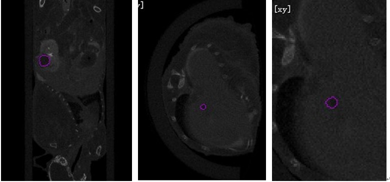
The results of Micro-CT scan reconstruction are shown in Figure 3. (a) is the tumor area in which the gastric cancer tumor metastasizes to the kidney (the area circled by the purple line), and (b) one of several metastases in the liver. Tumor tissue of micrometastases, (c) is a partial enlarged view of (b):
(a) (b) (c)
Figure 3 Contrast enhancement effect of tumor tissue and organ tissue by clinical iohexol contrast agent
Figure 4 Results of three-dimensional reconstruction after tumor and organ segmentation in nude mice
From FIG. 4 can clearly see the distribution of the tumor in mice, gastric cancer metastasis to three minute volume on liver tumor were 0.7767mm 3, 1.1125 mm 3, 1.1261mm 3, the tumor volume in the kidney It is 20.2666 mm 3 . The measured results after anatomy were consistent with the micro-CT scan results.
Experiment (3) Summary : The clinical use of iohexol contrast agent can also improve the contrast of organs or tumors in a certain way; this method of angiography can greatly save the expensive reagent cost of small animal contrast agents.
Conclusion: The ZKKS-MCT-III micro-CT system can be used to observe the growth and volume changes of tumor cells in mice in vivo, non-invasively and continuously. The experimental data show that not only can the instrument be used to monitor the modeling, but also the micro-CT system can accurately observe the tumor volume and shape during the treatment.
3D dental scanner is most commonly utilized for cosmetic and restoration purposes, such as reconstructive therapy or oral surgery. 3D imaging allows for planning and customizing services for procedures such as bone grafts, implants, and root canals. 3D scanning is also often used to create 3D images for orthodontic treatment.
Hand Held Dental 3D Scanner
Handheld 3D Scanner,3D Scanner Dental,Leica 3D Scanner,Android 3D Scanner
Shandong Carved Intelligent Technology Group Co.,Ltd , https://www.demetdent.com