2012 Cell Biology Technology Outlook
Although the methods and techniques for studying cell structure and function have made major breakthroughs, the pace of scientific exploration has never stopped. In 2012, perhaps we will see more imaging technologies appear, more fluorescent protein tools, ultra-high resolution imaging technology into new applications. In any case, every technological advancement in cell imaging methods will give us a deeper understanding of the inner world of cells. In 2008, a few years ago, cell imaging technology was named "Nature Methods" as the annual technology. After that, cell imaging technology has a new brilliance, higher resolution, faster and faster, not only can display ultra-high resolution images, but also quickly and sensitively capture the molecular motion in living tissue. In 2011, researchers at the University of California, San Francisco, reported a real-time imaging technique for living lung tissue—video-rate (two-photon imaging), which for the first time does not affect the normal physiological function of lung tissue. In the case of real-time imaging observations of cellular activities. Researchers from the US Department of Energy's Brookhaven National Laboratory have developed a new RatCAP PET live animal imaging system that provides neuroscientists with the ability to study brain function and behavior in animals that are awake and active. New tool. Although the methods and techniques for studying cell structure and function have made major breakthroughs, the pace of scientific exploration has never stopped. In 2012, perhaps we will see more imaging technologies appear, more fluorescent protein tools, ultra-high resolution imaging technology into new applications. In any case, every technological advancement in cell imaging methods will give us a deeper understanding of the inner world of cells. Ten years ago, the resolution of conventional optical microscopes stayed at 200 nm. Since then, ultra-high resolution imaging technology has emerged, and today optical microscopes can achieve resolutions of 20 nm. If you want to get a higher resolution, people have to use an electron microscope. Although the gap between the two imaging methods is getting smaller and smaller, it still exists, and biologists want to obtain the resolution of the electron microscope without fixing their samples. In 2012, we may see a wider application of electron microscopy technology. Biologists will connect the tools used in optics and electron microscopy to give people a deeper understanding of cells than ever before. In August 2011, Japanese scientist Atsushi Miyawaki and others reported a new reagent (Scale) in Nature Neuroscience. This magical reagent makes the biological sample optically transparent, but completely retains the fluorescent signal in the sample. As soon as the article was published, cell biologists and neurobiologists were excited, and the instrument company was equally excited. With the launch of this reagent, researchers are expected to delve into how neurons in the brain connect this year. In 2012, neurobiology will have a good show to be staged... In 2008, the discoverers and transformants of green fluorescent proteins were awarded the Nobel Prize in Chemistry, reflecting the importance of fluorescent proteins in cell biology research. As the understanding of the structure and function of fluorescent proteins deepens, researchers are constantly remodeling to develop new fluorescent proteins for new applications. In 2012, it is expected that the family of fluorescent proteins will continue to expand, and more new fluorescent proteins will be born, which are used in ultra-high resolution imaging, confocal microscopy, and even electron microscopy. Nowadays, understanding how cells respond to external forces and how the stiffness of the cellular microenvironment affects cell biology has also become a topic of interest to researchers. Atomic force microscopy (AFM) uses a microcantilever to sense and amplify the force between the sharp probe on the cantilever and the atom of the sample being tested for atomic resolution. Because the atomic force microscope can observe both the conductor and the non-conductor, it can make up for the shortcomings of the scanning tunneling microscope. With the increasing friendliness of AFM systems in recent years, new methods and techniques may emerge this year to explore how forces inside and outside the cell regulate everything, from cell movement to differentiation potential. (Source: BioTong website)
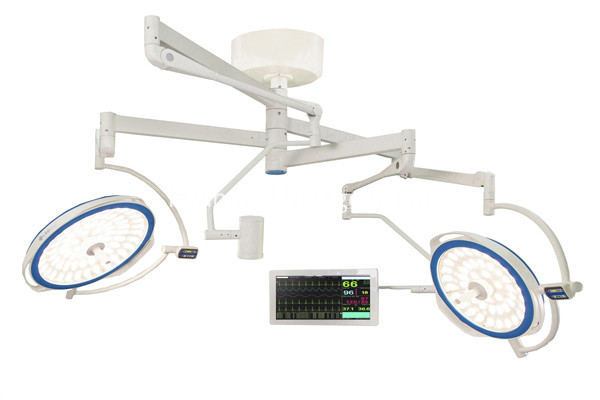
Round type sugical lamp is the most matured LED lamp in all LED series, it has producted for many years and exported to many countries, just like USA, UK, India, Italy, Thailand and other countries in the workd, almost zera maintainance rate, customers all give good feedback. the surgical lamp has the function of Clear lighting field; Outstanding color rendering index; Convenient removable sterilize handle; Control panel can choose touch screen control panel or normal control panel; it also has excellent color temperature adjustable technology; the operating lamp also can add HD camera system which is medical grade; for this OT lamp, it has ceiling type, wall type and mobile type; ceiling type can choose single dome, double dome or add camera system;
LED Surgery Lamp,LED Operating Lamp,LED Operating Light,LED Surgical Lamp Shandong Lewin Medical Equipment Co., Ltd. , https://www.lewinmed.com

